
Signalment and history

fig. 1 - Preoperative radiograph showing comminution of the proximal femoral metaphysis
A two–year-old, male, neutered DSH presented with a non-weightbearing lameness of the left pelvic limb, having been missing for most of the preceding week.
Initial investigations
Clinical examination was unremarkable other than crepitation of the affected limb. Radiology (fig. 1) revealed comminution of the proximal femoral metaphysis, to include the femoral neck. However, the articular surfaces of the left hip appeared intact. In addition, there was a small avulsion fracture of the caudal limit of the right ischium. Survey radiology of the chest and abdomen revealed no evidence of other injuries.
Treatment

fig. 2 - Postoperative radiograph showing 1/2-pins driven into ilium
The left femur was lengthened (by hanging leg preparation) and the stifle was aligned with respect to the hip joint, before the bone was stabilised in a closed manner with a combination of 2.4mm positive-threaded half-pins driven into the distal epiphysis and Ellis pins driven into the trochanteric region. The femoral connecting bar was then linked in a triangular fashion to pins driven into the left ilium and ischium (fig. 2). The frame was adjusted to maintain hip flexion at an approximate angle for weight bearing (see fig. 3) before all the clamps were tightened.
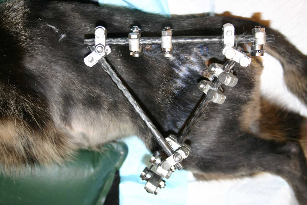
fig. 3 - triangulation of frame to fix hip in weight-bearing position
The cat was discharged the following day with instructions to confine him to a crate.
Follow up
The cat re-presented one month later and was able to walk reasonably well on the left pelvic limb; just using the stifle and hock joints. The frame was down-staged at this point to allow hip movement and cage rest was continued for a further three weeks. Radiology at eight weeks (fig. 4) revealed periosteal bridging and remodelling of the callus. All implants were removed at this point, and limited room exercise was allowed. Three months later, the owner reported good limb use with no
evidence of lameness.
Discussion
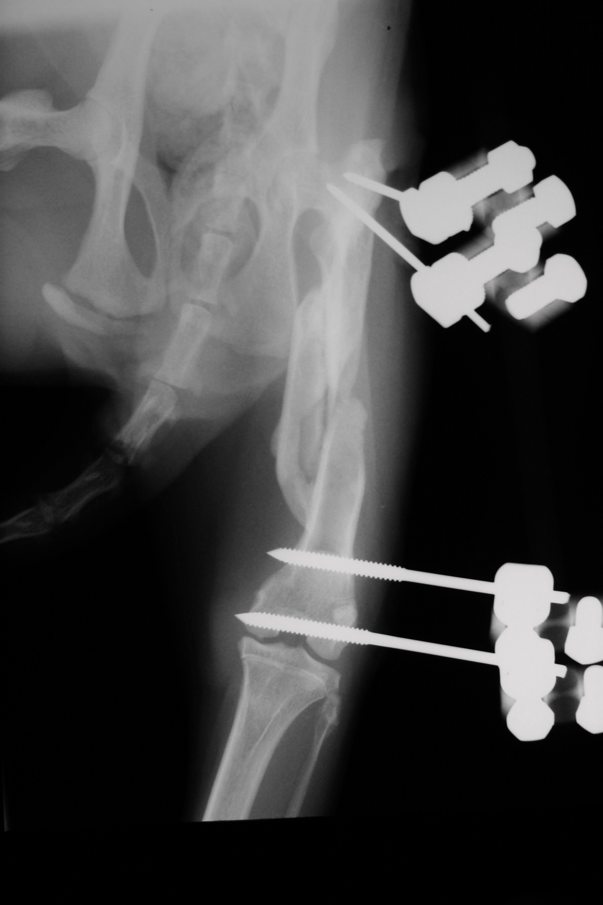
fig. 4 - Eight weeks post-op showing periosteal bridging
This case illustrates the principles of biological fracture healing; no effort is made to reconstruct the fragments and the callus is left undisturbed. The goals of surgery here are to re-establish limb length (although this is not critical as the joints will compensate to some degree for some shortening), ensure good joint alignment either side of the fracture and to provide rigid fixation to allow the callus to mineralise early.
Transarticular frames are very useful for managing fractures near a joint where bone-stock is limited. A degree of stiffness can be expected when a joint is immobilised for any length of time, but irreversible changes will not normally occur unless the joint is fixed for more than six-weeks.