
A 14-week-old female, entire Dachshund presented with a week-long history of seizures of increasing severity.
Investigations
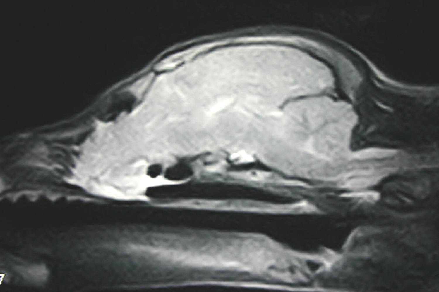
fig. 1 - sagittal T1W MR-scan of the skull, revealing rostral extension of neural tissue into nasal cavity
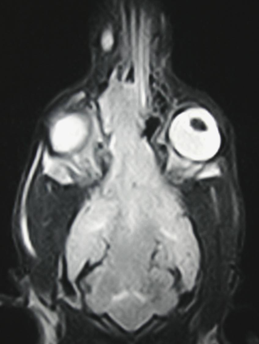
fig. 2 - Dorsal T1W MR-scan of the skull with neural tissue extending into the right side of the nasal cavity.
Neurological examination and blood tests, including serology, were unremarkable. MR-scans of the brain suggested abnormal development of the cranial vault, with extension of the brain rostrally into the nasal cavity (figs. 1 & 2, click to enlarge). Other differentials would include primitive neuroectodermal neoplasia and granuloma.
Further Investigations
Multiple Trucut biopsies were taken of the abnormal tissue via dorsal rhinotomy. The pup suffered several seizures within 24hrs of surgery, which responded to medical management.
Histopathology revealed normal neural tissue, consistent with a congenital anomaly of the skull.
Discussion
Although MRI was suggestive of the diagnosis, confirmation and therefore the ability to prognosticate sometimes requires biopsy. In this case, the site of the anomaly and therefore the approach was relatively straight forward, with a low risk of complications.
The rostral forebrain in general is very seizurogenic and the pup has since responded well to phenobarbitone adminstration, although she will probably require life-long therapy.